Glaucoma
Topics
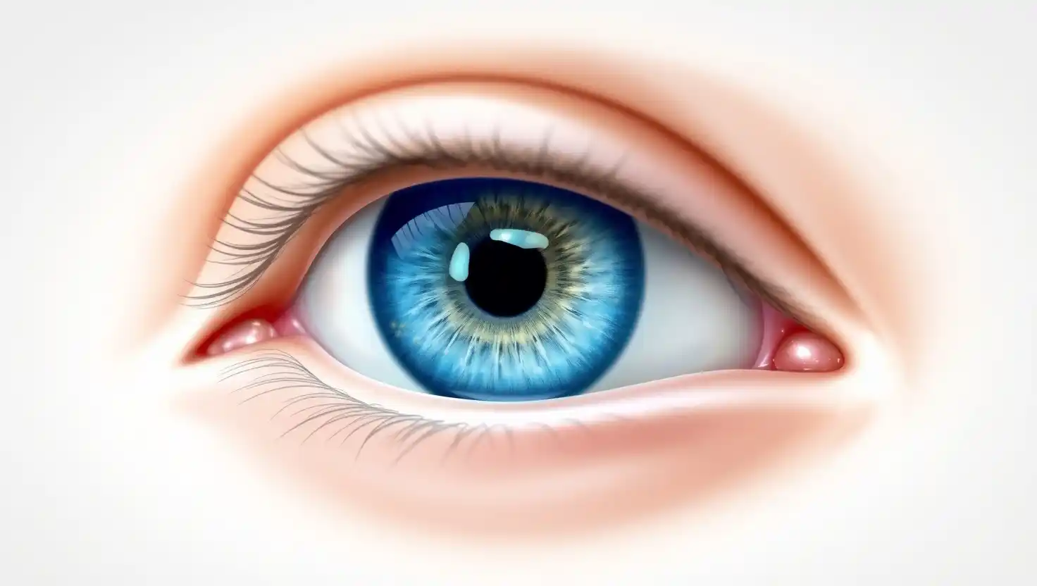
Introduction
Imagine gradually losing your ability to see the world around you, starting from the edges of your vision. This is often the insidious nature of glaucoma, a group of eye conditions that damage the optic nerve, the vital pathway that carries visual information from your eyes to your brain. Often referred to as the "silent thief of sight," glaucoma frequently progresses without noticeable symptoms in its early stages, making regular eye examinations crucial for timely detection and management.
What is Glaucoma?
Glaucoma is not a single disease but rather a group of progressive eye conditions that damage the optic nerve. This nerve is essential for sight, transmitting electrical impulses from the retina (the light-sensitive tissue at the back of the eye) to the brain, where these signals are interpreted as images. Damage to the optic nerve, often associated with an increase in the pressure inside the eye (intraocular pressure or IOP), can lead to gradual and irreversible vision loss.
The eye constantly produces a clear fluid called aqueous humour in the front part of the eye. This fluid nourishes the tissues and then drains out through a drainage angle at the junction of the iris (the coloured part of the eye) and the cornea (the clear front surface). If this drainage system becomes blocked or works too slowly, fluid can build up, increasing the IOP. Over time, this elevated pressure can damage the delicate nerve fibres of the optic nerve.
However, it's important to note that optic nerve damage and glaucoma can also occur in individuals with "normal" intraocular pressure, known as normal-tension glaucoma. In these cases, the optic nerve may be more sensitive to normal levels of pressure or there might be issues with blood flow to the nerve.
Regardless of the initial trigger, the hallmark of glaucoma is the progressive damage to the optic nerve, which initially affects peripheral (side) vision and can eventually lead to tunnel vision and, ultimately, blindness if not managed effectively.
Prevalence
Worldwide Prevalence: It is estimated that millions of people globally are living with glaucoma, and a significant proportion of them are unaware of their condition in the early stages. According to the World Health Organization (WHO), glaucoma is the second leading cause of blindness worldwide. Estimates suggest that over 70 million people globally have glaucoma, and this number is projected to increase in the coming years due to an ageing population.
Prevalence in India: India has a substantial burden of glaucoma. Studies indicate that glaucoma is a major cause of irreversible blindness in India, accounting for a significant percentage of all cases of blindness.
Types of Glaucoma
Glaucoma encompasses several distinct types, each with its own underlying mechanisms and characteristics:
Primary Open-Angle Glaucoma (POAG): This is the most common type of glaucoma worldwide. In POAG, the drainage angle of the eye appears open and normal, but the trabecular meshwork (the sieve-like structure through which aqueous humour drains) gradually becomes less efficient at draining fluid. This leads to a slow and progressive increase in intraocular pressure, damaging the optic nerve over time without noticeable early symptoms.
Angle-Closure Glaucoma (ACG) or Narrow-Angle Glaucoma: This type occurs when the iris blocks the drainage angle, preventing the outflow of aqueous humour. This blockage can happen suddenly (acute angle-closure), causing a rapid increase in IOP, severe eye pain, blurred vision, halos around lights, nausea, and vomiting – a medical emergency requiring immediate treatment. It can also develop gradually (chronic angle-closure), often without acute attacks.
Normal-Tension Glaucoma (NTG) or Low-Tension Glaucoma: In this type, optic nerve damage occurs despite the intraocular pressure being within the normal range. The exact causes are not fully understood, but it's believed that the optic nerve may be more sensitive to normal pressure levels or there might be problems with blood supply to the nerve.
Secondary Glaucoma: This type develops as a result of another eye condition or systemic problem. Causes can include eye injury, certain eye medications (especially steroid eye drops), eye tumours, inflammation (uveitis), advanced cataracts, or conditions like diabetes that affect blood vessels in the eye.
Congenital Glaucoma: This is a rare type of glaucoma that is present at birth or develops in early childhood. It is caused by abnormalities in the development of the eye's drainage system. Symptoms in infants may include cloudy eyes, excessive tearing, and unusual sensitivity to light.
Causes of Glaucoma
While the exact causes of primary glaucoma are not fully understood, several factors are known to contribute to the development of the different types:
Increased Intraocular Pressure (IOP): In many forms of glaucoma, elevated IOP is a significant risk factor. This increase can be due to an imbalance between the production and drainage of aqueous humour. In POAG, the drainage system becomes less efficient over time. In ACG, the iris physically blocks the drainage angle.
Optic Nerve Sensitivity: In normal-tension glaucoma, the optic nerve may be inherently more susceptible to damage even at normal IOP levels. Factors like reduced blood flow to the optic nerve are also thought to play a role.
Genetics: There is evidence to suggest that genetics play a role in the development of glaucoma, particularly POAG and congenital glaucoma. Having a family history of glaucoma increases an individual's risk.
Secondary Conditions: As mentioned earlier, secondary glaucoma is caused by other eye conditions or systemic problems that affect the eye's drainage system or overall health.
Eye Injury or Surgery: Trauma to the eye or certain eye surgeries can sometimes lead to secondary glaucoma.
Medications: Prolonged use of certain medications, especially steroid eye drops, can increase IOP and the risk of secondary glaucoma.
Blood Flow Issues: Problems with blood circulation to the optic nerve may contribute to optic nerve damage, particularly in normal-tension glaucoma.
Symptoms of Glaucoma
One of the most challenging aspects of glaucoma, particularly primary open-angle glaucoma, is that it often progresses without noticeable symptoms in its early stages. Vision loss typically begins in the peripheral (side) vision and develops gradually. Many people do not realise they have glaucoma until significant optic nerve damage has occurred and they start experiencing noticeable vision loss.
Symptoms can vary depending on the type of glaucoma:
Primary Open-Angle Glaucoma (POAG):
- Gradual loss of peripheral (side) vision, often in both eyes.
- In advanced stages, tunnel vision (the ability to see only straight ahead).
Acute Angle-Closure Glaucoma: This type presents with sudden and severe symptoms, including:
- Severe eye pain.
- Blurred vision.
- Halos (rainbow-coloured circles) around lights.
- Nausea and vomiting.
- Redness of the eye.
- A hazy cornea.
- This is a medical emergency requiring immediate attention.
Normal-Tension Glaucoma (NTG):
- Similar to POAG, often involves gradual loss of peripheral vision.
Secondary Glaucoma: Symptoms can vary depending on the underlying cause. They may overlap with POAG or ACG symptoms.
Congenital Glaucoma: Symptoms in infants may include:
- Cloudy eyes.
- Excessive tearing (epiphora).
- Unusual sensitivity to light (photophobia).
- Larger than normal eyes (buphthalmos).
Due to the often silent progression of POAG, regular comprehensive eye examinations that include testing of the optic nerve and visual fields are essential for early detection before significant vision loss occurs.
Diagnosis of Glaucoma
Diagnosing glaucoma involves a comprehensive eye examination and a series of tests to assess the health of your optic nerve and the pressure inside your eye. Your ophthalmologist will typically perform the following:
- Tonometry: This test measures the intraocular pressure (IOP). Different methods can be used, including the "puff of air" test (non-contact tonometry) and Goldmann applanation tonometry (which requires numbing eye drops and gentle contact with the eye).
- Ophthalmoscopy: This allows the doctor to examine the optic nerve directly. They will look for signs of damage, such as cupping (an enlargement of the central depression in the optic disc).
- Visual Field Testing (Perimetry): This test maps your peripheral (side) vision to identify any areas of vision loss, which is a characteristic sign of glaucoma damage. You will be asked to look straight ahead and indicate when you see small lights appearing in different parts of your visual field.
- Gonioscopy: This test helps determine the type of glaucoma by examining the drainage angle of the eye (where the iris meets the cornea). A special lens and a slit lamp are used to visualise this area and check if the angle is open or blocked.
- Optical Coherence Tomography (OCT): This imaging technique uses light waves to create detailed cross-sectional images of the optic nerve and retinal nerve fibre layer. It helps to measure the thickness of these layers and detect early signs of damage.
- Pachymetry: This test measures the thickness of the cornea. Corneal thickness can influence IOP measurements, so this information is important for accurate assessment.
The diagnosis of glaucoma is based on a combination of these test results, along with your medical history and a thorough clinical examination. Regular monitoring with these tests is also crucial to track the progression of the disease and the effectiveness of treatment.
Treatment of Glaucoma
The primary goal of glaucoma treatment is to lower intraocular pressure (IOP) to a level that prevents or slows down further damage to the optic nerve and preserves vision. The specific treatment approach depends on the type and severity of glaucoma. Common treatment options include:
Eye Drops (Medications): These are the most common initial treatment for glaucoma. Various types of eye drops are available, each working in a different way to reduce IOP, either by decreasing the production of aqueous humour or by increasing its outflow. Examples include prostaglandin analogues, beta-blockers, alpha-adrenergic agonists, and carbonic anhydrase inhibitors. Often, more than one type of eye drop may be needed. It's crucial to use these medications regularly as prescribed by your doctor. (Brand available:Combigan)
Laser Treatment: Several types of laser procedures can be used to treat glaucoma:
- Selective Laser Trabeculoplasty (SLT): This laser targets specific cells in the trabecular meshwork to improve drainage in open-angle glaucoma. It can often be repeated if needed.
- Argon Laser Trabeculoplasty (ALT): Similar to SLT, but uses a different type of laser. It is also used for open-angle glaucoma.
- Laser Peripheral Iridotomy (LPI): This procedure creates a small hole in the iris to improve fluid flow and prevent or treat angle-closure glaucoma.
- Cyclophotocoagulation: This laser treatment targets the ciliary body (which produces aqueous humour) to reduce fluid production. It is often used in more advanced cases of glaucoma.
Surgery: If eye drops and laser treatment are not sufficient to control IOP or if the glaucoma is advanced, surgery may be necessary:
- Trabeculectomy: This is a traditional surgical procedure where the surgeon creates a new drainage channel for fluid to leave the eye, forming a small bleb (fluid-filled blister) under the upper eyelid.
- Minimally Invasive Glaucoma Surgery (MIGS): These newer surgical techniques use tiny devices to enhance the eye's natural drainage pathways with less invasive procedures compared to trabeculectomy. Examples include iStent, Hydrus Microstent, and XEN Gel Stent. MIGS procedures often have a faster recovery time and fewer complications.
- Drainage Implants (Tube Shunts): A small tube is inserted into the eye to drain fluid to a reservoir placed under the conjunctiva (the clear outer layer of the eye). This is often used in complex or refractory cases of glaucoma.
Treatment for glaucoma is usually lifelong and requires regular follow-up appointments with your ophthalmologist to monitor your IOP, optic nerve health, and visual fields, and to adjust your treatment plan as needed.
Risk Factors
- Risk of glaucoma rises significantly after age 60.
- A family history of glaucoma increases your likelihood of developing it.
- High intraocular pressure is a key risk factor for glaucoma.
- People of African and Asian descent are at higher risk for certain types of glaucoma.
- Conditions like diabetes, high blood pressure, heart disease, and sickle cell anaemia elevate glaucoma risk.
- Eye injuries can lead to secondary glaucoma.
- Long-term corticosteroid use can raise eye pressure and glaucoma risk.
- A thin central cornea may increase your susceptibility to glaucoma.
- Structural abnormalities in the optic nerve head can heighten glaucoma risk.
- Severe short-sightedness (high myopia) is linked to a greater risk of open-angle glaucoma.
Complications
- Glaucoma causes progressive vision loss, starting with peripheral vision.
- If untreated, glaucoma can lead to irreversible blindness.
- Vision loss from glaucoma can drastically reduce independence and quality of life.
Tips to Live with Glaucoma
Living with glaucoma requires proactive management and adaptation. Here are some tips to help you maintain your quality of life:
- Adhere to Your Treatment Plan: Use your eye drops regularly and as prescribed. Consistency is key to controlling IOP.
- Communicate with Your Doctor: Report any changes in your vision or any difficulties you are having with your treatment.
- Organise Your Medications: Use pillboxes or reminders to ensure you don't miss any doses of your eye drops.
- Optimise Your Home Environment: Improve lighting, reduce clutter, and eliminate tripping hazards to enhance safety, especially as your vision changes.
- Utilise Low Vision Aids: If you experience vision loss, explore low vision aids such as magnifiers, large-print materials, and adaptive technology.
- Stay Active and Healthy: Regular exercise and a balanced diet can contribute to overall well-being.
- Plan for Transportation: If your vision loss affects your ability to drive safely, make alternative transportation arrangements.
- Protect Your Remaining Vision: Wear sunglasses to protect your eyes from UV radiation and consider using side shields on your glasses.
- Stay Positive: While living with glaucoma can be challenging, maintaining a positive outlook and focusing on what you can still do can improve your overall well-being.
Common Misconceptions About This Condition
Misconception: Glaucoma has obvious early symptoms.
Reality: Primary open-angle glaucoma, the most common type, typically has no noticeable symptoms in its early stages. This is why it's often called the "silent thief of sight."
Misconception: If my vision is good, I don't have glaucoma.
Reality: You can have significant optic nerve damage from glaucoma and still have good central vision, especially in the early stages. Peripheral vision is usually affected first.
Misconception: Glaucoma is just about high eye pressure.
Reality: While high intraocular pressure is a major risk factor, glaucoma can also occur in people with normal eye pressure (normal-tension glaucoma). The damage is to the optic nerve, regardless of the pressure level.
When to See a Doctor
You should see an ophthalmologist or optometrist for a comprehensive eye examination if you:
- Have a family history of glaucoma.
- Are over the age of 40 (or earlier if you have risk factors).
- Experience any changes in your vision, such as blurred vision, halos around lights, or loss of peripheral vision.
- Have had an eye injury.
- Use steroid eye drops regularly.
- Have certain medical conditions like diabetes or high blood pressure.
- Experience sudden, severe eye pain, redness, nausea, and vomiting (which could be acute angle-closure glaucoma – a medical emergency).
Questions to Ask Your Doctor
When you visit your doctor for glaucoma or a routine eye exam, consider asking the following questions:
- What is my eye pressure? Is it in a healthy range?
- How does my optic nerve look? Are there any signs of damage?
- What are my risk factors for glaucoma?
- How often should I have my eyes examined?
- Do I have glaucoma? If so, what type?
- What is the stage of my glaucoma?
- What are my treatment options?
- What are the potential side effects of the recommended treatments?
- How often and how should I use my eye drops?
How to Support Someone Dealing with Glaucoma
Supporting someone with glaucoma involves understanding the challenges they face and offering practical and emotional support:
- Offer Practical Assistance: Help with tasks that become difficult due to vision loss, such as reading small print, navigating unfamiliar environments, or organising medications.
- Provide Transportation: If their vision affects their ability to drive, offer rides to appointments or other necessary places.
- Encourage Adherence to Treatment: Gently remind them to use their eye drops and attend their appointments.
- Help Adapt Their Environment: Assist in making their home safer and more accessible by improving lighting, reducing clutter, and addressing tripping hazards.
- Accompany Them to Appointments: Offer to go with them to doctor's appointments for support and to help take notes or ask questions.
Conclusion
Glaucoma is a serious eye condition that can lead to irreversible vision loss if left undetected and untreated. Due to its often silent progression, regular comprehensive eye examinations are paramount for early diagnosis and management. While there is currently no cure, consistent adherence to prescribed treatments, regular monitoring, and adopting helpful lifestyle adjustments can significantly slow the progression of the disease and maintain a good quality of life.
Frequently Asked Questions
Can glaucoma be cured?
Is glaucoma hereditary?
Can I prevent glaucoma?
What is the normal eye pressure?
How often should I get my eyes checked for glaucoma?
Download the app


DawaaDost
Our Policies
Our Services
Knowledge Base
Fetured Categories


© 2026 DawaaDost. All rights reserved. In compliance with Drugs and Cosmetics Act, 1940 and Drugs and Cosmetics Rules, 1945, we don't process requests for Schedule X and other habit forming drugs.